Inside a Feather 1 was presented at the Society for Integrative and Comparative Biology annual meeting 2017, in New Orleans. It introduces the work we present here (Inside a Feather II). Both posters are available to view here:
Both posters are on the subject of elliptical voids inside the feather cortex.
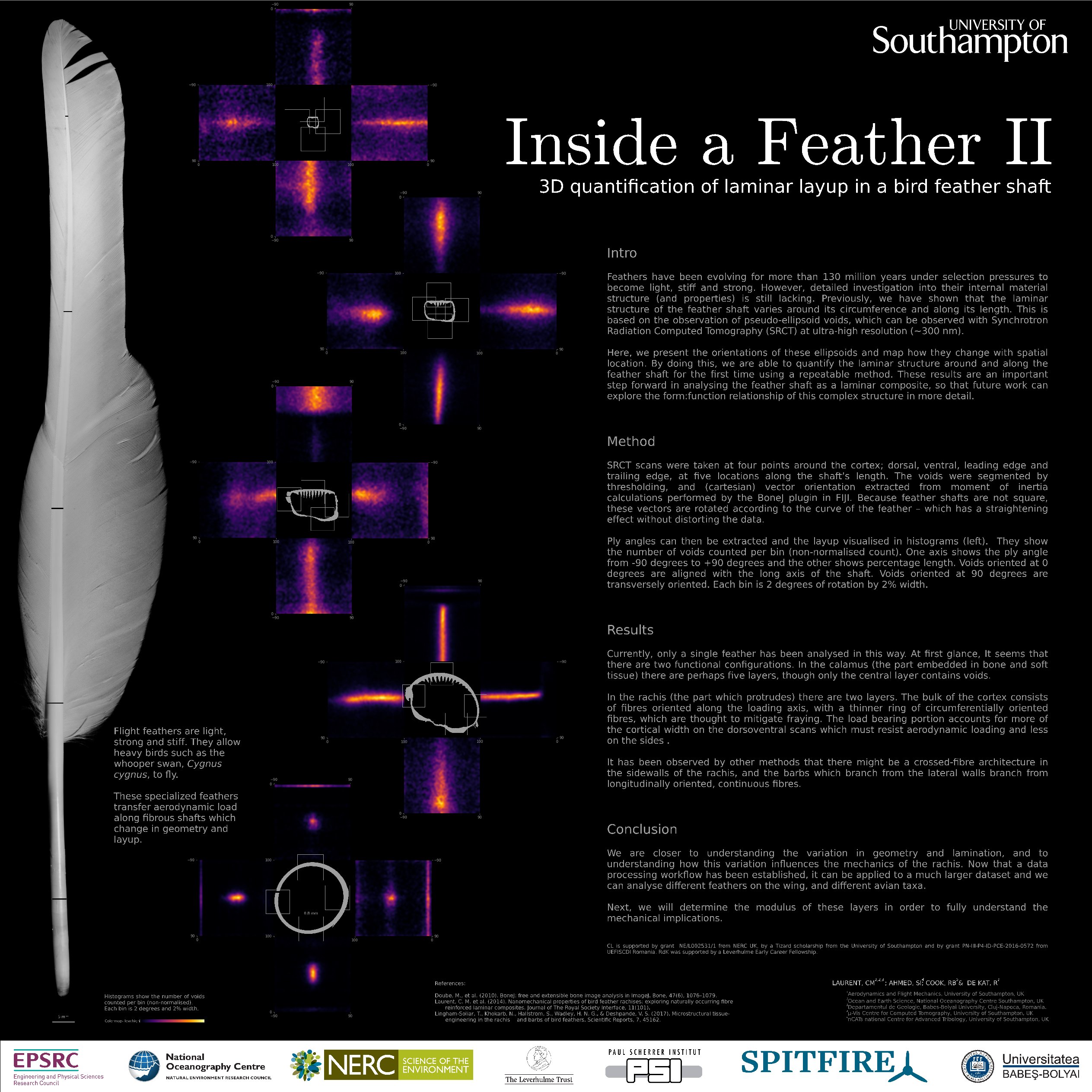
In this poster, we present the orientation of these inter-fibre voids, but unfortunately there is not enough space to show them on the poster, and it's difficult to illustrate them in a meaningful way in two dimensions - so here they are in three!
Below, there are some 3D representations of reconstructed CT data. Click once, and they should be loaded in a 3D viewer (this might take a few seconds).
...you can orbit around the data and zoom with the usual touchscreen gestures on your smart device.
... then click the  button in the pop up menu on the bottom right corner and enjoy stereoscopic virtual reality. Be careful your phone's backlight isn't set to autodim after a few seconds - you will need to wake it up again.
You may also teleport yourself around this virtual world by looking at the floor (circular cross-hairs) and pressing the silver 'cardboard button'. Be careful not to go too far, though, or you might get lost. If you do - just hit refresh :)